What you need to know about a Head Ultrasound
Contents
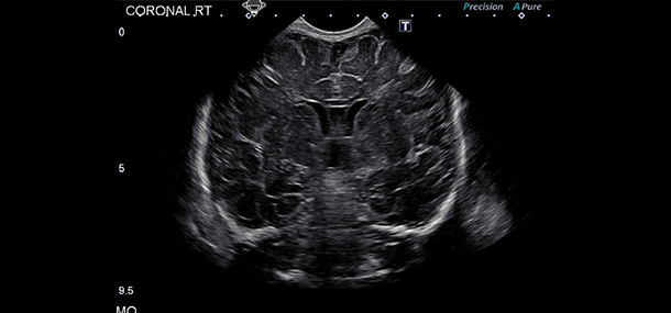
A head ultrasound is a test that uses sound waves to make images of the brain and cerebrospinal fluid. This test is usually carried out when there is a concern about neurological problems in an infant. Premature babies or babies that are born more than 3 weeks before the due date, usually have a head ultrasound to check for brain problems that can occur, such as:
-
Injury to the white matter of the brain that surrounds the ventricles (periventricular leukomalacia or PVL).
-
Bleeding in the brain (intraventricular hemorrhage or IVH)
Doctors may also order an head ultrasound for babies with:
-
A bulging in the soft spot of the head
-
An abnormal increase in head size
-
Any symptoms of nerve or brain problems.
The test can also help doctors diagnose other problems of the brain, such as:
-
Infection
-
Suspected complications of meningitis
-
Cysts, tumors, or other masses
-
Too much fluid in the brain or ventricles (hydrocephalus).
Doctors most often use head ultrasound on infants younger than 6 months old because infants have a soft spot on top of their heads where the skull has not yet grown together. The gap allows the sound waves through. It’s rarely done in older children and adults because of the bones of their skull block sound waves. However, adults may need a head ultrasound to find a tumor or mass during brain surgery.

What does the Procedure Involve?
During a head ultrasound, your baby will be positioned on their back or stomach. If necessary, your baby can also be in your arms when the procedure is carried out.
A technician (sonographer) will start the procedure by spreading a clear gel on your baby’s scalp, over the soft spot of the head (fontanel). The gel can help the transmission of the sound waves. Then, the technician will gently move a small wand-like device (transducer) over the gel. High-frequency sound waves are emitted by the device and how the sound waves bounce back from your baby’s head is measured by a computer. The sound waves are changed into images by the computer. Once the sonographer is done, they will clean the gel off of your baby’s head.
The whole procedure is painless, but your child might feel a little cold and wet from the gel, as well as slight pressure on the head as the transducer is moved. Sometimes, babies cry in the ultrasound room, particularly if they are restrained. However, this will not interfere with the procedure. You can offer your baby’s favorite toy or a pacifier, or even feed them for comfort.
In adults who need a head ultrasound to find a tumor or mass during brain surgery, the procedure is only done after the skull has been opened because the sound waves cannot pass through the skull.
How Long Should I Stay in the Area?
Your baby can leave the hospital right away after the head ultrasound but plan to stay in the area for about two days because the results are usually ready within 1 to 2 days. Once the results are ready, you will have to attend a follow-up appointment with your child’s doctor to discuss the results. If you are an adult who needs head ultrasound as part of brain tumor surgery, you will need to stay in the hospital for about 3 to 10 days and stay in the local area for 14 additional days.
What’s the Recovery Time?
There is no recovery time after a head ultrasound. Your child can be back to their usual behavior immediately. For head ultrasound as part of brain surgery, the recovery time depends on the reason you have to undergo the surgery.
What About Aftercare?
No special aftercare is needed after a head ultrasound and you can feed your baby as you normally do.
What’s the Success Rate?
Head ultrasound is an effective procedure with high success rates. The procedure is found to be 100% correct in identifying patients with germinal matrix hemorrhage and 93% accurate in identifying patients who don’t have the condition.
Head ultrasound is also a very safe procedure. Since radiation is not involved in head ultrasound, no risks are associated with this test.
Are there Alternatives to Head Ultrasound?
Below are some of the alternatives to head ultrasound:
-
Magnetic resonance imaging (MRI) of the brain – this test uses a magnetic field and radio waves to create images of the brain and brain stem. In some cases, this test can provide clear images of parts of the brain that cannot be seen clearly with a head ultrasound.
-
Computed tomography (CT) scan of the head – during this test, a special X-ray machine is used to take pictures of your child’s brain, skull, sinuses, and blood vessels. However, since it uses radiation, there are some risks associated with this procedure.
What Should You Expect Before and After the Procedure
Before a head ultrasound, your child’s doctor may need to ensure that no problem occurs in your child’s brain because your child is born prematurely or shows symptoms of brain or nerve problems. After the procedure, the results of the test should help the doctor make an accurate diagnosis. If any problem or abnormality is found, the doctor can create a treatment plan or order other tests to confirm.
For an in-depth analysis of a Head Ultrasound Procedure, watch this short video.
https://youtube.com/watch?v=HYOmwLtzCAo
To check prices or to book a Head Ultrasound Procedure, in Thailand or anywhere else in the world, head on over to MyMediTravel now!

